


Introduction
The incidence of chronic subdural hematoma (CSH) is estimated to be 1.7 to 20.6 per 100,000 persons per year, and it is expected to increase with the advent of an aging society, as it mostly occurs in the elderly population [1]. Head trauma is a well-known cause of CSH; however, in the elderly after the age of 65 years, there may not be any history of trauma [2,3]. This can be explained by the occurrence of cerebral atrophy in the elderly and increased use of anti-thrombotic drugs.
Numerous studies have been published on other factors that contribute to the development of CSH, such as chronic alcoholism, liver disease, and malignancy, which are known to affect coagulopathy [2,4-8]. The most commonly accepted pathophysiological explanation of CSH is that trivial head trauma leads to tearing of bridging veins, which causes abundant bleeding and leads to the development of a hematoma [5,9]. However, another study has focused on several key courses involved in CSH development, which include inflammation, angiogenesis, and fibrinolysis because trauma may be absent of very trivial and does not explain the developmental course of CSH [10].
Specifically, first, inflammation occurs after damage to the dural border cells, and then angiogenesis occurs in the membrane, causing growth of fragile and leaky vessels [4,10-12]. Blood leakage and fluid exudation occur from these vessels, and at the same time, a fibrinolytic process occurs to prevent clot formation. Through these processes, neomembrane growth and hematoma growth continue to occur, leading to CSH development [4,10-12]. Also, in the presence of cerebral atrophy, a bridging vein is present in the subdural space, which has a more fragile and thin vessel wall than the subarachnoid space [13]. Therefore, tearing of the bridging vein can occur even without any trauma. Cerebral atrophy occurs mainly in the elderly population, and, an increasing number of elderly patients take anti-thrombotic drugs, which can affect the bleeding tendency due to medical conditions.
There are several mechanisms of anti-thrombotic drugs. Aspirin selectively inhibits cyclooxygenase-1, resulting in dysfunction of platelets. Clopidogrel and prasugrel irreversibly inhibit the P2Y12 receptor and patients need to take these drugs once a day. Unlike thienopyridines, ticagrelor does not bind to the adenosine diphosphate binding site and instead reversibly binds to a distinct site on the P2Y12 receptor and patients need to take this drug twice a day. Cilostazol inhibits phosphodiesterase III; thus, inhibiting platelet aggregation. Warfarin is a vitamin K-dependent oral anticoagulant that inhibits the coagulation process, Non-vitamin K antagonist oral anticoagulants inhibit factor Xa, and dabigatran inhibits factor IIa to inhibit the coagulation process. Each anticoagulant drug has a different half-life and bioavailability [14].
Although the clear mechanism of CSH is not known, CSH shows several density types in computed tomography (CT) findings, and each type has different mechanisms. Previous studies have reported the predictive factors for recurrence in terms of both clinical and radiological factors preoperatively, and one of the factors is anti-thrombotic drugs [15,16]. There are no previous reports on the relationships between anti-thrombotic drugs and the possibility of operation in CSH. In this study, we investigated the association between anti-thrombotic drugs and the possibility of operation in CSH with consideration of both clinical and radiological factors.
Material and Method
This study was conducted after receiving approval from the Institutional Review Board (IRB) of Seoul Medical Center (IRB No. 2022-02-008-001), and all patients were recruited from a single institution. We reviewed all patients who were diagnosed with CSH at our institution between January 2011 and December 2021. All diagnoses of CSH were confirmed on the basis of a CT scan. We excluded data obtained from 47 patients who had previously undergone brain surgery and 47 patients who refused treatment. As a result, we retrospectively analyzed 254 patients who were admitted to our institution to receive treatment.
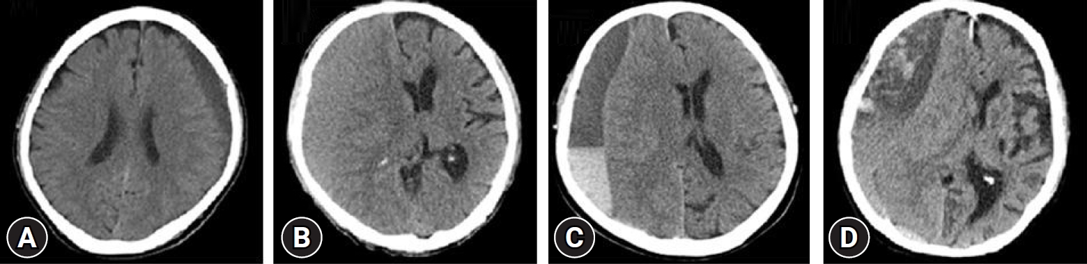
We obtained factors, such as gender, age, hypertension (HTN), diabetes mellitus (DM), hyperlipidemia, liver disease, chronic alcoholism, cerebrovascular disease (CVD), coronary heart disease (CHD), malignancy, and anti-thrombotic drugs, from the past medical records. All patients stopped taking anti-thrombotic drugs after the diagnosis of CSH. The following initial laboratory findings related to bleeding tendency were investigated: platelet count, prothrombin time (PT) percentage, and international normalized rate (INR). Clinical findings, such as initial Glasgow coma scale (GCS) and history of trauma, were also investigated. Patients diagnosed with CSH on initial or follow-up CT examination were treated based on the Markwalder grading system (Table 1) [17], in which patients with a score of 2 to 4 were treated surgically and those with a score of 0 to 1 received conservative treatment. The following initial CT findings were investigated: initial maximal thickness of subdural hematoma, density type of CSH, location of CSH (bilateral, left, or right), and distance of midline shifting. Density type of CSH was categorized into the following 4 types: homogeneous hypodensity (<25 Housefield unit [HU]), homogeneous isodensity (25-35 HU), mixed type, and layered type on CT findings (Fig. 1). We defined the mixed type as a heterogeneous subdural hematoma and the layered type as a subdural hematoma of different stages separated by a layer. We performed mostly one burr hole trephination with or without irrigation and in layered and mixed type of CSH, we performed craniotomy with hematoma removal.
We defined recurrence as a case of reoperation due to an increase in the subdural hematoma volume in the ipsilateral subdural space seen on CT within 6 months postoperatively, and the decision about reoperation was based on the Markwalder grading system.
Statistical analysis: Data are presented as the mean±standard deviation, and number is used (percentage) for categorical variables. Categorical variables were assessed using Pearson χ2 and Fisher exact tests to examine the presence of an association between the variables and the possibility of operation in CSH. Differences were considered statistically significant if P-values were <0.05. IBM SPSS ver. 26.0 (IBM Corp., Armonk, NY, USA) was used for statistical analyses.
Results
Demographic characteristics
The baseline characteristics are presented in Table 2. The mean age was 75.45±8.25 years (60-99 years). There were 167 men and 87 women in this study. Past medical history of HTN, DM, hyperlipidemia, liver disease, chronic alcoholism, CHD, CVD, and malignancy was present in 94 (37.0%), 84 (33.1%), 53 (20.9%), 7 (2.8%), 52 (20.5%), 54 (21.3%), 42 (16.5%), and 19 (7.5%) patients, respectively. Medication with anti-thrombotic drugs included history of antiplatelet drugs in 58 patients (22.8%), history of anticoagulant drugs in 14 patients (5.5%), and none of the 2 medications in 182 patients (71.7%). A total of 145 patients (57.1%) had a history of trauma. Initial laboratory findings showed a mean INR of 1.0911±0.3165 (range, 0.81-4.67), a mean PT percentage of 94.26±19.710 (range, 16-154), and a mean platelet count of 228.14±97.633 (range, 40-667). An initial GCS score of 15 was noted in 98 patients (38.6%), an initial GCS score of 13 to 14 was noted in 47 patients (18.5%), an initial GCS score of 8 to 12 was noted in 107 patients (42.1%), and an initial GCS score of 3 to 7 was noted in 2 patients (0.8%). The authors conducted one burr hole trephination in 123 cases (76.3%) with or without irrigation and in layered and mixed type of CSH, we performed craniotomy with hematoma removal in 38 cases (23.6%).
CT findings of CSH
The mean hematoma thickness in CT findings was 15.66±8.74 mm. The maximal thickness of hematoma was 45 mm, and the minimal thickness of hematoma was 3 mm. The most common hematoma density type was the mixed density in 92 patients (36.2%), followed by isodensity in 79 patients (31.1%), hypodensity in 57 patients (22.4%), and layered density in 26 patients (10.2%). The mean distance of midline shifting in CT findings was 6.34±8.22 mm. The maximal distance of midline shifting was 55 mm, and the minimal distance of midline shifting was 0 mm. The most common laterality of CSH was the right side in 96 patients (37.8%), followed by the left side in 85 patients (33.5%) and bilaterally in 70 patients (27.6%).
Factors affecting the possibility of operation of CSH
Summary of the baseline characteristics and the possibility of operation in 254 patients with CSH are described in Table 3. We compared 2 groups; the no operation group (n=93, 36.6%, Markwalder grading score 0 to 1) and the operation group (n=161, 63.3%, Markwalder grading score 2 to 4), to investigate which factors were associated with the possibility of operation. There was no significant association between age and the possibility of operation (P=0.742). Also, there was no significant association between gender and the possibility of operation (P=0.388). There were no significant associations between all comorbidities and the possibility of operation. Prior medication was not significantly associated with the possibility of operation (P=0.159). Initial laboratory findings, such as INR (P=0.593), PT percentage (P=0.272), and platelet count (P=0.526), were not significantly associated with the possibility of operation. Head trauma was significantly associated with the possibility of no operation (P=0.002). Initial GCS was significantly associated with the possibility of operation (P<0.05). With respect to radiologic findings, hematoma thickness, density type, midline shifting, and laterality were significantly associated with the possibility of operation (P<0.05). Mixed type had the highest possibility of operation in CSH (n=69, 42.9%), but the hypodensity type had the lowest possibility of operation in CSH (n=13, 8.1%). Right side had the highest possibility of operation in CSH (n=72, 44.7%), but bilaterality had the lowest possibility of operation in CSH (n=30, 18.6%).
Anti-thrombotic drugs with head trauma or chronic alcoholism
We analyzed the associations between anti-thrombotic drugs and the possibility of operation in patients with trauma or anti-thrombotic drugs and the possibility of operation in patients with chronic alcoholism in Table 4. In patients with head trauma, 30 of 80 patients with an operation history (37.5%) had taken anti-thrombotic drugs, whereas 12 of 65 patients without any operation history (18.4%) had taken anti-thrombotic drugs. In patients with chronic alcoholism, 26 of 37 patients with an operation history (70.2%) had taken anti-thrombotic drugs, whereas 4 of 15 patients without any operation history (26.6%) had taken anti-thrombotic drugs. Medication with anti-thrombotic drugs was statistically associated with the possibility of operation, especially in head trauma (P=0.043) or chronic alcoholism groups (P=0.018).
Anti-thrombotic drugs associated with CSH recurrence
We found a significant association between the density type and anti-thrombotic drugs (P=0.037) in Table 5. The most prominent density type associated with anti-thrombotic drugs was the mixed type (n=37, 40.2%). We found that the use of anti-thrombotic drugs was significantly associated with the recurrence of CSH (P=0.021) in Table 6.
Discussion
Aging and increase use of anti-thrombotic drugs
In recent years, the incidence of CHD and CVD has been increasing in the elderly population, and the use of anti-thrombotic drugs has been increasing accordingly [18]. CSH is one of the complications of anti-thrombotic drugs, and several studies have reported a correlation between the use of anti-thrombotic drugs and the incidence of CSH [4,18]. Like recent trends, this study showed that the mean age of the patients was 75.45 years, and 28.3% of them were taking anti-thrombotic drugs.
Anti-thrombotic drugs in patients with head trauma
Previous studies have reported a significant association between anti-thrombotic drugs and prevalence of CSH [2,19-23]. In this study, medication with anti-thrombotic drugs was not significantly associated with the possibility of operation in CSH (P=0.159). This can be explained by the fact that inhibition of the clot formation process due to medication with anti-thrombotic drugs was not significantly associated with the degree of hematoma expansion to the extent of requiring operative treatment. Also, head trauma was significantly associated with the possibility of no operation in CSH (P=0.002). However, in this study, we found that the use of anti-thrombotic drugs was significantly associated with the possibility of operation in patients with a head trauma history (P=0.043). This suggests that medication with anti-thrombotic drugs has a more significant effect on the expansion of hematoma, which was adequate enough to require operative treatment than head trauma. Thus, we suggest that close observation might be needed if patients diagnosed with CSH took anti-thrombotic drugs with a head trauma history. There was no significant relationship between the possibility of operation and medication with anti-thrombotic drugs in the patient group with no head trauma history (P=0.926).
Anti-thrombotic drugs in patients with chronic alcoholism
Previous studies have reported a significant association between chronic alcoholism and prevalence of CSH [1,8,10]. It is known that chronic alcoholism can affect the bleeding tendency because it can cause thrombocytopenia, and the Framingham Heart Study suggested a strong association between alcoholism and brain atrophy [24,25]. In this study, chronic alcoholism was not significantly associated with the possibility of operation in CSH (P=0.192). However, we found that the medication with anti-thrombotic drugs was significantly associated with the possibility of operation in patients with chronic alcoholism (P=0.019). This can be explained by the fact that alcohol-induced thrombocytopenia alone is not significantly associated with the degree of hematoma expansion to the extent of requiring operative treatment; instead, the combination of inhibition of clot formation caused by anti-thrombotic drugs and thrombocytopenia caused by chronic alcoholism could cause hematoma expansion to the extent of requiring operative treatment. This process can also be explained by considering that patients with chronic alcoholism could have accompanying cerebral atrophy. Thus, we suggest that close observation might be needed if patients diagnosed with CSH had a history of chronic alcoholism and anti-thrombotic drug medication. There was no significant association between the possibility of operation and medication with anti-thrombotic drugs in the patient group without chronic alcoholism (P=0.357).
CT findings in patients with use of anti-thrombotic drugs
The isodensity type was often found to be the most common than the other density types in previous studies [26,27]. In this study, the isodensity type was found to be the second most common type (n=79, 31.1%) after the mixed type (n=90, 35.4%). Other types included the layered type (n=26, 10.2%) and the hypodensity type (n=57, 22.4%). The mechanism of the mixed type could be explained by accumulation of repeated microbleeding [26]. Repeated trauma may also result in acute subdural hematoma in CSH. Also, due to the different stages of subdural hematomas with a history of trivial trauma, a chronic-on-CSH may lead to the mixed density [28]. The authors speculated that the use of anti-thrombotic drugs makes increase in the risk of mixed type of CSH and can be associated with the possibility of operation. However, another study reported that there was no significant association between medication with anti-thrombotic drugs and density type in CT and magnetic resonance imaging (MRI) findings [29]. It can be explained by the fact that in some cases, CSH with a homogeneous density on CT shows mixed density on Diffusion weighted MRI [29]; however, we were only gathering information from CT findings. Therefore, a prospective study with MRI findings as well as CT findings is needed to investigate which factors are associated with the possibility of operation in CSH.
Recurrence of CSH
The recurrence rate of CSH varies from study to study, and in this study, it was 45.9% (74 of 162). A previous report showed that the presence of initial symptoms, such as headache, DM, prior anticoagulant medication, and postoperative midline shifting, are independent predictors for recurrence of CSH [16]. In addition, other studies have shown that the presence of brain atrophy is associated with recurrence because of the presence of a persistent subdural space [15,30]. Furthermore, if there is septation in radiologic findings, recurrence may be caused by a residual hematoma because it is difficult to drain the hematoma completely [29,31,32]. Also, if there is a residual hematoma, a neovascularized membrane develops, and therefore, continuous microbleeding in fragile and leaky vessels can occur, which can lead to hematoma accumulation. In other words, recurrence can occur if the hematoma continues to grow and there is a capacity of the subdural space for such a hematoma to accumulate sufficiently. Many studies have been performed to assess several factors that can affect recurrence [16,30,33,34]. In this study, the association between the use of anti-thrombotic drugs and postoperative recurrence was significant (P=0.021). In this study, anti-thrombotic drugs were stopped immediately after the diagnosis of CSH, and the half-life and offset and onset of each drug were different. The effectiveness of anti-thrombotic drugs can still be residual, which can also affect the hematoma volume, and it is different for each agent. Therefore, through a prospective study, it is necessary to investigate the association between the onset of repeated medication after the initial operation and recurrence.
This study has some limitations. This was a retrospective study, and therefore, it was subjected to potential sources of bias. Although we showed statistical significance in the subgroup study, the number of subgroups was not very large; thus, a prospective study with a larger number of data is needed.
Conclusion
This study reports that the initial GCS and CT findings, such as hematoma thickness, density type, midline shifting, and laterality were significantly associated with the possibility of operation in CSH. Furthermore, in patients taking anti-thrombotic drugs, head trauma or chronic alcoholism was significantly associated with the possibility of operation and close monitoring of these patients should be considered in those situations.
 PDF Links
 PubReader
 ePub Link
 Full text via DOI
 Download Citation
 Print
Print


