J Korean Soc Geriatr Neurosurg > Volume 18(1); 2022 > Article
Cho, Oh, and Kim: Intramedullary schwannoma at the thoracic level in a geriatric patient: a case report

Abstract

Intramedullary schwannoma, a disease that primarily occurs in middle-aged individuals, is a very rare tumor that occurs inside the spinal cord, constituting only 0.3% of spinal cord tumors. These tumors have most often been reported to involve a single lesion in the spinal cord. A magnetic resonance imaging (MRI) examination and histopathological report can confirm the diagnosis of intramedullary schwannoma. We describe a case of intramedullary schwannoma in a 71-year-old female patient. The tumor was evaluated using MRI and through a pathophysiological assessment. The lesion showed diffuse bulbous thickening with an ill-defined central signal change of the spinal cord from C6 to T6 on T2-weighted MRI. During surgery, a reddish and adhesive mass was removed. After the operation, the patient's leg weakness improved. Despite the rarity of the disease, we should consider intramedullary schwannoma as a possible tumor in the differential diagnosis of intramedullary lesions in elderly patients, for which surgical treatment should be considered.

Introduction

Schwannomas occur most frequently within the intradural-extramedullary compartment. However, intramedullary schwannoma is a very rare intraspinal cord tumor. It accounts for only 1.1% of spinal schwannoma [1]. It is a disease that occurs in commonly in middle age [2]. However, this case has clinical significance in that it occurred in an elderly person.
We report a rare case of thoracic intramedullary schwannomas in elderly explored by magnetic resonance imaging (MRI) and pathogenesis within the spinal cord.

Case Report

A 71-year-old female patient was admitted to our hospital with gradual paraparesis with urinary difficulty for 10 days. She had no previous spinal disease, nor any traumatic history.
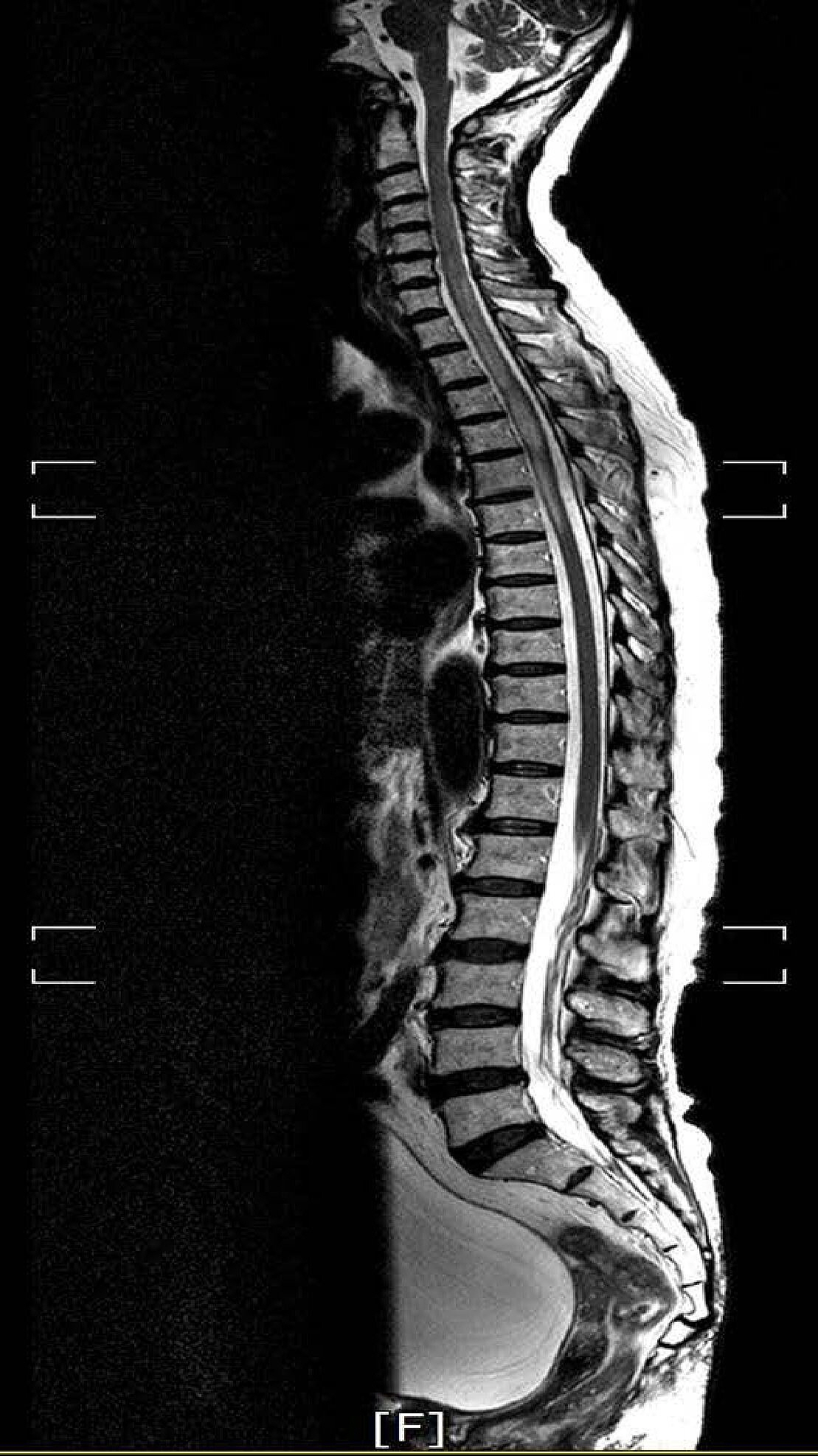
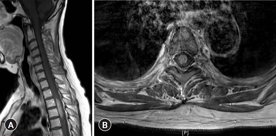
Neurological examination revealed that the motor power of the patient’s left leg was grade IV and right leg GIII. The sensation of all modalities under T5 dermatomes was reduced. Her bilateral knee jerks and ankle jerks were hyperactive and well sustained ankle clonus was observed. The lesion showed diffuse bulbous thickening with ill-defined central signal change of spinal cord from C6 to T6 level on the T2 weight-image of MRI (Fig. 1). An ovoid, well-marginated enhanced mass was located at the center of the cord on the gadolinium enhanced image (Fig. 2).
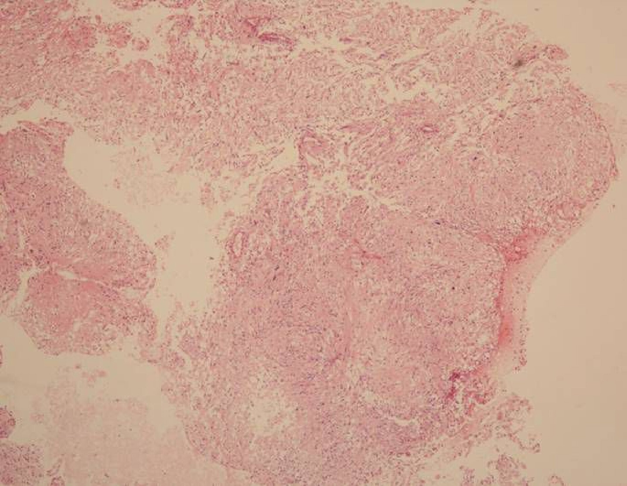
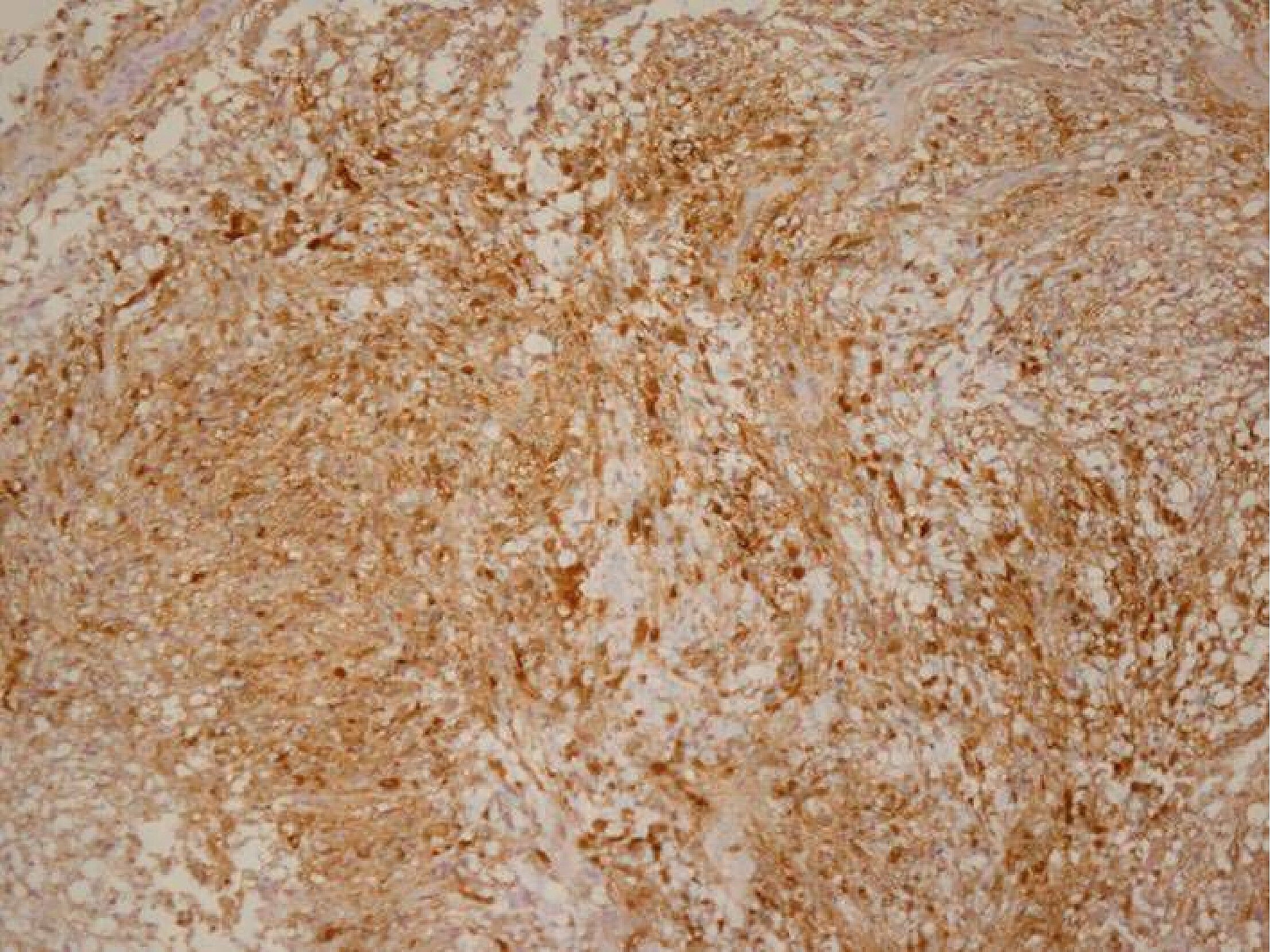
The operation was performed with standard posterior midline approach. After laminoplasty from T2 to T5, a midline dura incision with lateral retraction was performed. Midline myelotomy showed reddish and some infiltrative adhesive mass lesion. Frozen section of the lesion was defined as a schwannoma. Due to the infiltrative adhesive nature, its total resection was not easy. During the procedure, the intraoperative monitoring was uneventful. After the operation, the patient’s weakness was improved, and IV+ and self-voiding was possible. Histological analysis confirmed the diagnosis of schwannoma (Figs. 3-5).

Discussion

James Kernohan has been recognized as the first neurosurgeon to report an intramedullary schwannoma case in 1952. However, Penfield had already reported an intramedullary tumor with schwannomas characteristics in 1932 [3].
Intramedullary schwannoma is a rare disease among non- neurofibroma patients, accounting for 0.3% of intraspinal schwannomas and 1.1% of spinal schwannoma [4]. The rarity of intramedullary schwannoma is due to the absence of Schwann cells in the central nerve system including the parenchyma of the brain and spinal cord, thus the pathogenesis of intramedullary schwannoma is still controversial. None of specific theories has gained acceptance. There are 5 hypotheses: (1) the intramedullary perivascular nervous plexus along Schwann cells, (2) the migration of Schwann cells during embryonic development, (3) the aberrant intramedullary nerve fibers related to Schwann cells, (4) neoplastic growth of Schwann cells in dorsal root entering zone, and (5) focal intramedullary proliferation of Schwann cells in reaction to chronic diseases or trauma [5-7].
The ratio of male to female of intramedullary schwannomas is 3:1. The mean age of disease presentation was 40.2 years, middle age [2]. The common region of intramedullary schwannomas is the cervical spine (63%), the thoracic spine (26%), and the lumbar (11%) levels [8].
MRI is one of the choices for the diagnosis of spinal cord tumors. Intramedullary Schwannoma is often known to be hypointense on T1 weighted image, and hyperintense on T2 weighted image [9]. However, signal intensity depends on macroscopic features and histological composition such as cysts, hemorrhage, and necrosis [5]. Thus diagnostic MRI of the intramedullary schwannoma is very limited and pathological analysis is important for diagnosis.
Common pathological finding is Antoni type A or mixture with Antoni type B. Schwannoma cell cytoplasm was reactive to S-100 protein but non-reactive to glial fibrillary acidic protein and epithelial membrane antigen [10].
The main treatment of schwannoma is surgical excision. Complete removal of tumor is usually possible and prognosis of disease is satisfactory [7,11].

Conclusion

Despite the rarity of intramedullary schwannoma it should be considered as one of the possible tumors in intramedullary lesion. Although this this disease is common in middle-aged people, it is a disease that should require differential diagnosis even in geriatric patients. The main treatment of the disease is its surgically total excision; however, its adhesive nature makes the removal difficult. It is essential to recognize the adhesive nature of the intramedullary schwannoma.

NOTES

Conflicts of interest

No potential conflict of interest relevant to this article was reported.

Acknowledgments

We thank the patient for her valuable participation in this study.

Fig. 1.
A T2 weighted sagittal image showing diffuse bulbous thickening with ill-defined central signal change of the spinal cord, from C6 to T6. Written informed consent was obtained for publication of this case report and accompanying images.
jksgn-2022-00010f1.jpg
Fig. 2.
An approximately 35-mm ovoid, well-marginated mass with enhancement in the spinal cord at T3 to T4. (A) Sagittal image, (B) axial image. Written informed consent was obtained for publication of this case report and accompanying images.
jksgn-2022-00010f2.jpg
Fig. 3.
Microscopic image showing a biphasic pattern with cellular Antoni A and hypocellular Antoni B areas (H&E, ×40). Written informed consent was obtained for publication of this case report and accompanying images.
jksgn-2022-00010f3.jpg
Fig. 4.
Microscopic image revealing compact fasciles of elongated tumor cells with slight nuclear polymorphism (H&E, ×100). Written informed consent was obtained for publication of this case report and accompanying images.
jksgn-2022-00010f4.jpg
Fig. 5.
Immunohistochemical stains demonstrating diffuse positivity for S-100 protein (×100). Written informed consent was obtained for publication of this case report and accompanying images.
jksgn-2022-00010f5.jpg

REFERENCES

1. Brown KM, Dean A, Sharr MM. Thoracic intramedullary schwannoma. Neuropathol Appl Neurobiol 2002;28:421-4.
crossref pmid
2. Swiatek VM, Stein KP, Cukaz HB, et al. Spinal intramedullary schwannomas-report of a case and extensive review of the literature. Neurosurg Rev 2021;44:1833-52.
crossref pmid pmc pdf
3. Penfield W. Notes on operative technic in neurosurgery. Ann Surg 1946;124:383-5.
crossref pmc
4. Hayashi F, Sakai T, Sairyo K, et al. Intramedullary schwannoma with calcification of the epiconus. Spine J 2009;9:e19-23.
crossref
5. Yang T, Wu L, Deng X, Yang C, Xu Y. Clinical features and surgical outcomes of intramedullary schwannomas. Acta Neurochir (Wien) 2014;156:1789-97.
crossref pmid pdf
6. Herregodts P, Vloeberghs M, Schmedding E, Goossens A, Stadnik T, D’Haens J. Solitary dorsal intramedullary schwannoma: case report. J Neurosurg 1991;74:816-20.
crossref pmid
7. Kim SD, Nakagawa H, Mizuno J, Inoue T. Thoracic subpial intramedullary schwannoma involving a ventral nerve root: a case report and review of the literature. Surg Neurol 2005;63:389-93.
crossref pmid
8. Nicácio JM, Rodrigues JC, Galles MH, Faquini IV, de Brito Pereira CA, Ganau M. Cervical intramedullary schwannoma: a case report and review of the literature. Rare Tumors 2009;1:e44.
crossref pmid pmc
9. Riffaud L, Morandi X, Massengo S, Carsin-Nicol B, Heresbach N, Guegan Y. MRI of intramedullary spinal schwannomas: case report and review of the literature. Neuroradiology 2000;42:275-9.
crossref pmid pdf
10. Kahilogullari G, Aydin Z, Ayten M, Attar A, Erdem A. Schwannoma of the conus medullaris. J Clin Neurosci 2005;12:80-1.
crossref pmid
11. Ryu KS, Lee KY, Lee HJ, Park CK. Thoracic intramedullary schwannoma accompanying by extramedullary beads-like daughter schwanommas. J Korean Neurosurg Soc 2011;49:302-4.
crossref pmid pmc


ABOUT
BROWSE ARTICLES
EDITORIAL POLICY
FOR CONTRIBUTORS
Editorial Office
21, Namdong-daero 774beon-gil, Namdong-gu, Incheon 21565, Korea
Tel: +82-32-460-3304    Fax: +82-32-460-3899    E-mail: editor@jksgn.org                

Copyright © 2024 by Korean Society of Geriatric Neurosurgery.

Developed in M2PI

Close layer
prev next